

The Relationship Between Smoking and Mental Illness
According to a study by the American Centers for Disease Control and Prevention, Bargain E-Juice says Americans with various mental illnesses are more likely to smoke than regular people by 70 percent. Aside from this, with vaping being a new trend, more people could actually be at risk.
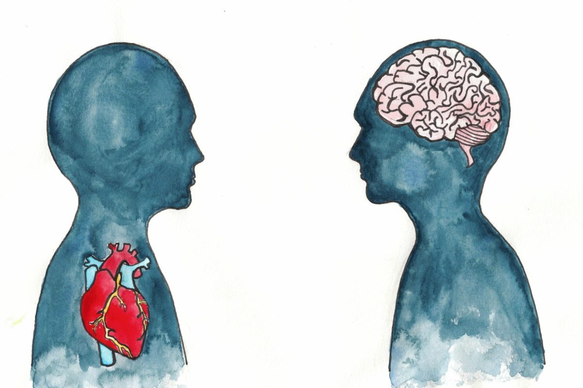
As a result, psychologists are hard at work trying to understand this situation and help their patients to quit. Some of the severe illnesses that have been linked to tobacco usage include heart disease and lung cancer.
However, further complicating the situation is that people who are suffering from …